
Could you recognize a possible skin cancer growth on your skin? Clinical studies have shown that even the most experienced physicians can accurately diagnose 71% of malignant lesions and 94% of benign lesions. This strongly suggests that biopsies are indicated since even experienced physicians are unable to predict malignancy. Even so, the first defense you have against skin cancer is recognizing changes in your skin early, so you and your doctor can address them before they become a problem, if possible.
The ABCD Checklist
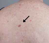
We all have freckles, birth marks, moles and other skin irregularities somewhere on our bodies. Most of these are harmless. However, it is important to get to know your skin and all of its unique marks. Precancerous and cancerous skin lesions often look a little different from benign spots.
The ABCD checklist has been reported to have a high likelihood of detecting melanoma. Use the following guidelines to assess an area of your skin, and if you notice any of the following, it’s best to see your dermatologist for further evaluation:
- A=asymmetry
- B=border irregularity
- C=color variation
- D=diameter of greater than 6mm (about 1/4 inch)
However, if you are worried about any lesion, regardless of size or appearance, or you notice recent changes, such as increased size, alteration of shape or color, itching, burning, bleeding or pain, see your dermatologist. It is better to be proactive about skin cancer.
Types of Tumors
Not all skin lesions are cancerous, but it can be difficult to know the difference by just looking at the skin. The following pictures will help illustrate the different types of benign and malignant lesions that dermatologists and plastic surgeons can address.
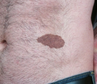
Benign Lesions
The most common pigmented lesions are benign nevi: junctional, compound and intradermal. They can mimic melanoma along with other benign lesions such as seborrheic keratoses and solar lentigines.
Pre-cancerous – atypical, dysplastic or Clark’s nevi may all lead to melanoma. Actinic keratosis may transform to either basal or squamous cell cancer. Trichoepithelioma may mimic basal cell cancer. Keratoacanthoma may closely mimic squamous cell cancer.
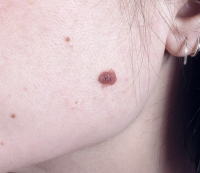
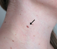
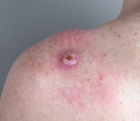
Malignant Lesions
Sometimes skin cancer can look very similar to a benign lesion; other times it looks quite different. The most common types of skin cancers Dr. Parker treats in our practice are basal cell, squamous cell or malignant melanomas.
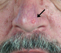
Basal cell carcinoma is the most common type of skin cancer, and it can usually be completely removed when treated early. Basal cell cancer typically does not spread quickly, but it can run deep beneath the skin, so treatment is very important.
Squamous cell carcinoma is most commonly seen on skin after many years of sun damage, but it can occur in any patient. It spreads more easily than basal cell carcinoma, but can usually be cured if caught early enough, before it spreads to the lymph nodes. Early detection is key.
Melanoma is the most serious of skin cancers. It spreads very quickly, and if it spreads more deeply into the skin or other tissues, melanoma can be fatal. The good news is, if it is caught early enough, while it is still in the top layers of skin, melanoma can be successfully removed and the patient be cured. Again, early detection is crucial for the patient’s health.
Skin Cancer Treatment
Skin cancer can be treated in a number of different ways, depending on the severity of the lesion. You may seek treatment from a dermatologist, plastic surgeon, or both. Typically, larger lesions will be treated by a plastic surgeon, who is trained to restore the area to a more normal appearance using reconstructive skin grafts, or flaps.
Benign lesions
These lesions can be treated in a variety of ways depending on the discussions between the treating physician and patient. Because these lesions are benign and are very unlikely to become cancerous, watching or observing them over time is a very logical course of action.
Patients who wish to have them removed for cosmetic or other reasons may elect to do so by one of two ways. A dermatologist will often remove these lesions by shaving or scraping them down to the second level of the skin, called the dermis. Plastic surgeons will often remove these lesions by excising, or cutting them out, and carefully repairing the wound with stitches.
The ultimate appearance after one of these procedures varies based on the size and location of the lesion, type of skin, age of the patient and skill of the practitioner. The patient should understand that some type of scar will almost always be present. It is very important for patient and doctor to discuss how conspicuous this scar will likely be in order for the patient to make an appropriate informed decision.
Precancerous lesions
These lesions should usually be biopsied to obtain a tissue diagnosis. Actinic keratoses can be treated topically but atypical, dysplastic, Clark’s nevi, trichepiteliomas, and keratoacanthomas are almost always removed. Dermatologists can remove some of these lesions if they are deemed small enough. Larger lesions are usually referred to a plastic surgeon for excision.
The ultimate appearance after one of these procedures varies based on the size and location of the lesion, type of skin, age of the patient and skill of the practitioner. The patient should understand that some type of scar will almost always be present. It is very important for patient and doctor to discuss how conspicuous this scar will likely be. Because these lesions will often transform into skin cancers, removal is usually recommended, however.
Malignant lesions
These lesions are always biopsied with a definitive tissue diagnosis prior to removal. This is important because the biopsy will tell not only what type of skin cancer is present, but also its level of aggressiveness. Options for removal are based on the type of cancer, level of aggressiveness, size and location of the lesion, and overall medical health of the patient.
These options include the following:
- Removal by a dermatologist with cryotherapy or electrodessication and curettage for superficial cancers on the trunk or extremities; more recently topical treatment has been used by some dermatologists for superficial cancers.
- Surgical removal by a plastic surgeon or specially trained dermatologists, known as a Moh’s surgeon.
- Radiation therapy may be employed by a radiation oncologist in certain circumstances.
Leave a Reply